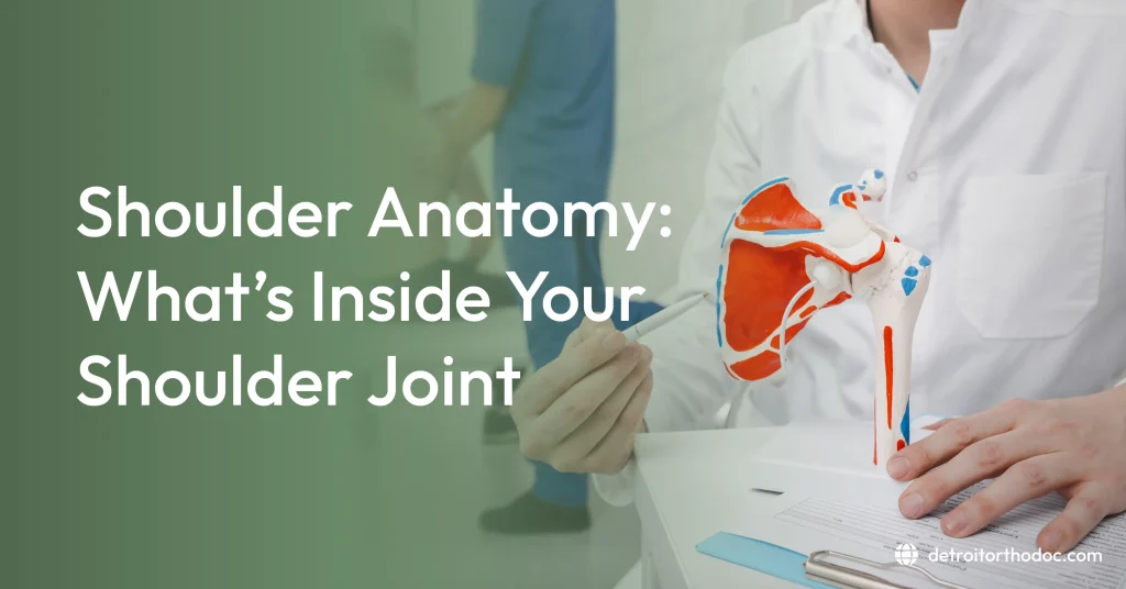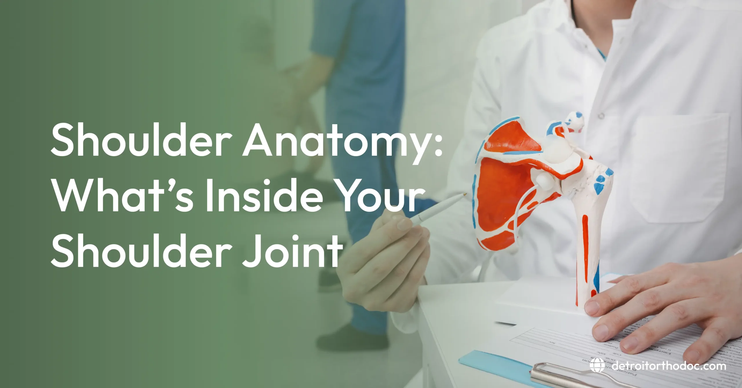
Shoulder anatomy consists of three bones (the clavicle, scapula, and humerus), four joints (glenohumeral, acromioclavicular, sternoclavicular, and scapulothoracic), and the soft tissues that hold them together: the labrum, joint capsule, ligaments, rotator cuff muscles and tendons, and bursae. This design gives the shoulder the widest range of motion in the body, and it is also why the shoulder is injured so often.
The shoulder is the most mobile joint in the human body, and understanding shoulder anatomy explains both why that is true and why the shoulder is injured so often. Reaching overhead, throwing, brushing your hair, and putting on a coat all depend on a structure that trades stability for freedom of movement.
That trade-off is built into the design. The head of the upper arm bone is roughly three to four times larger than the shallow socket it sits against, meaning only about a third of the ball contacts the socket at any moment (Kenhub). The glenohumeral joint is described in clinical literature as the most mobile and least stable joint in the body, and the most commonly dislocated (NIH StatPearls).
This guide walks through the anatomy of the shoulder from the outside in. It starts with the four joints that work together, then covers the three bones that form them, and then looks inside the joint itself at the labrum, capsule, ligaments, cartilage, and bursae.
You will also find sections on the muscles, the nerves that supply the shoulder, how stability is maintained, normal range of motion, and which structures are involved in common injuries. Understanding these parts makes it easier to follow a diagnosis, ask better questions, and understand why a particular treatment is recommended.
Shoulder Joint Anatomy: A Complex of Four Joints
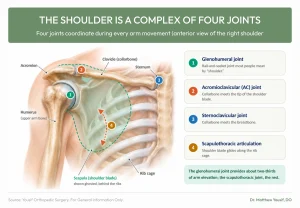
One of the most useful things to understand about human shoulder anatomy is that the shoulder is not one joint. It is a complex of four joints that coordinate with each other during every arm movement. The clavicle and scapula together form the shoulder girdle, which links the arm to the trunk.
The four joints are:
- Glenohumeral joint, the ball-and-socket joint most people mean when they say shoulder.
- Acromioclavicular joint, where the collarbone meets the tip of the shoulder blade.
- Sternoclavicular joint, where the collarbone meets the breastbone.
- Scapulothoracic articulation, where the shoulder blade glides along the rib cage.
During arm elevation, the glenohumeral joint provides roughly two-thirds of the motion, with the scapulothoracic articulation contributing the remaining third (NIH StatPearls). This is why shoulder blade movement matters so much, and why problems with scapular control often produce pain at the joint itself.
Shoulder Bone Anatomy: The Bones of the Shoulder
Three bones form the structure of the shoulder, and they are simpler than the soft tissue around them. The bones of the shoulder are:
- Clavicle (collarbone), the strut connecting the arm to the chest and the only bony link between the upper limb and the axial skeleton.
- Scapula (shoulder blade), a flat triangular bone that provides attachment for most shoulder muscles.
- Humerus (upper arm bone), whose rounded head forms the ball of the joint.
The humeral head also has two bony prominences, the greater and lesser tuberosities, which serve as attachment points for the rotator cuff tendons. A groove between them houses the long head of the biceps tendon. These landmarks matter clinically, since fractures and tendon problems are often described by their relationship to them.
Shoulder Blade Anatomy
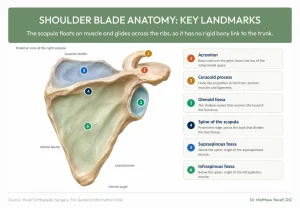
The scapula is the foundation of the whole complex. It has no rigid bony attachment to the trunk, instead floating on muscle and gliding across the ribs.
Key features of the scapula include:
- Glenoid fossa, the shallow socket that receives the humeral head.
- Acromion, the bony roof over the top of the joint.
- Coracoid process, a hook-like projection at the front serving as a muscle and ligament anchor.
- Spine of the scapula, the prominent ridge across the back.
- Supraspinous and infraspinous fossae, the surfaces above and below that ridge where rotator cuff muscles originate.
The acromion is clinically significant because the space beneath it, the subacromial space, is where rotator cuff tendons pass. Narrowing of this space contributes to shoulder impingement, one of the most common sources of shoulder pain.
Inside the Joint: The Glenoid Labrum
When people ask about the anatomy of the shoulder joint, the labrum is often the structure they have heard about without quite understanding. It is a ring of fibrocartilage attached around the rim of the socket. The cross-section below shows how it sits alongside the capsule, cartilage, bursa, and rotator cuff tendon.
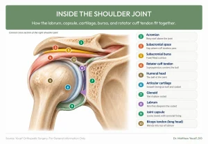
The labrum serves several purposes:
- It deepens the glenoid socket, contributing up to half of the socket’s total depth (Medscape).
- It provides an attachment point for the glenohumeral ligaments.
- It helps create a seal that contributes to joint stability.
- At its upper portion, it is continuous with the long head of the biceps tendon.
That final point explains why labral tears at the top of the socket, known as SLAP tears, often involve the biceps tendon as well. Labral injuries typically follow dislocations, falls onto an outstretched arm, or repetitive overhead throwing, and they can cause clicking, catching, or a sense of instability. You can read more about shoulder labral tears and related conditions.
The Joint Capsule and Shoulder Ligaments
The anatomy of the human shoulder joint includes a fibrous capsule that surrounds the entire joint like a loose sleeve. It is lined internally by a synovial membrane that produces lubricating fluid.
The capsule is deliberately loose, which permits the shoulder’s enormous range of motion. Thickened bands within it form the ligaments of the shoulder, which provide passive restraint. Ligaments connect bone to bone, while tendons connect muscle to bone; the shoulder tendons are covered in the muscle section below.
- Superior, middle, and inferior glenohumeral ligaments, which tighten in different arm positions to limit excessive movement.
- Coracohumeral ligament, running from the coracoid process to the humerus.
- Coracoacromial ligament, forming part of the protective arch above the joint.
- Coracoclavicular ligaments, the trapezoid and conoid, which stabilize the acromioclavicular joint.
The capsule is central to frozen shoulder, where inflammation and thickening cause it to contract. That is why frozen shoulder produces genuine loss of motion even when someone else moves your arm.
Cartilage and Bursae
Two more structures inside and around the joint deserve mention, since both are common sources of pain.
Articular cartilage
A smooth layer of articular cartilage covers the humeral head and the glenoid surface, allowing them to glide against each other with minimal friction. When this cartilage wears down, the result is shoulder osteoarthritis, producing pain, stiffness, and sometimes grinding.
Bursae
Bursae are small fluid-filled sacs that reduce friction where tendons pass over bone. The shoulder contains several, and three are worth knowing, with the first mattering most clinically:
- Subacromial bursa, sitting between the rotator cuff tendons and the acromion above.
- Subscapular bursa, between the subscapularis tendon and the glenoid neck.
- Subcoracoid bursa, between the coracoid process and the subscapularis.
Inflammation of the subacromial bursa, called subacromial bursitis, frequently accompanies rotator cuff problems and produces pain with overhead reaching. Figure 3 above shows where the subacromial bursa and the subacromial space sit above the rotator cuff tendon.
Anterior Shoulder Anatomy
The front of the joint is clinically important because most shoulder dislocations occur in the forward direction. Structures at the front include:
- Subscapularis, the rotator cuff muscle on the front surface of the shoulder blade.
- Long head of the biceps tendon, running through its groove into the joint.
- Coracoid process and the muscles attaching to it.
- Anterior capsule and glenohumeral ligaments, the primary restraints against forward instability.
- Anterior labrum, frequently torn during dislocation.
A tear of the anterior labrum following dislocation is called a Bankart lesion. Because the anterior capsule and labrum are the main barriers to the humeral head sliding forward, damage here often leads to recurrent instability, particularly in younger patients.
Posterior Shoulder Anatomy
The back of the shoulder handles outward rotation and helps slow the arm during throwing. Back shoulder anatomy includes:
- Infraspinatus and teres minor, the rotator cuff muscles producing external rotation.
- Posterior deltoid, pulling the arm backward.
- Posterior capsule, which can become tight in overhead athletes.
- Posterior labrum, less commonly injured than the anterior labrum.
Posterior capsule tightness is a recognized issue in throwing athletes and can alter shoulder mechanics. The back of the shoulder is also where the scapular stabilizing muscles do their work, positioning the socket so the ball can move efficiently.
Shoulder Muscle Anatomy
Shoulder muscle anatomy is a large subject, so this section covers the essentials. Muscles are the shoulder’s primary stabilizers, compensating for a bony structure that offers little inherent security (NIH StatPearls, Shoulder Muscles). The main groups are:
- Rotator cuff, four deep muscles that center the humeral head in the socket: the supraspinatus, infraspinatus, teres minor, and subscapularis.
- Deltoid, the large outer muscle producing powerful elevation in all directions.
- Scapular stabilizers, including the trapezius, rhomboids, serratus anterior, and levator scapulae.
- Large movers, including the pectoralis major and latissimus dorsi.
The biceps and triceps also cross the shoulder and contribute to its function, which is where the anatomy of arm and shoulder muscles overlaps. The rotator cuff tendons attach the cuff muscles to the humeral head, and these small muscles do the essential work of keeping the ball centered while the larger muscles generate movement (NIH StatPearls, Rotator Cuff). When these tendons are injured, the result is a rotator cuff tear.
Nerves and Blood Supply
The shoulder is supplied by nerves arising from the brachial plexus, a network formed by nerve roots exiting the neck. Because that network begins in the neck, neck and shoulder anatomy are closely linked, and a problem in the neck can cause pain that is felt in the shoulder. Two nerves are especially relevant clinically:
- Axillary nerve, which powers the deltoid and provides sensation over the outer shoulder. It is routinely checked after a dislocation because of its vulnerable position.
- Suprascapular nerve, which supplies the supraspinatus and infraspinatus.
Blood supply comes primarily from branches of the axillary artery, including the anterior and posterior circumflex humeral arteries. This anatomy explains why numbness over the outer shoulder after an injury is a meaningful finding, and why nerve assessment is part of any thorough shoulder examination.
How the Shoulder Stays Stable
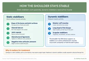
Given how little bony support exists, stability is an active achievement rather than a structural given. Clinicians divide the stabilizers into two categories.
Static stabilizers work without muscle activity:
- The shape of the bones and the joint surfaces
- The glenoid labrum
- The joint capsule
- The glenohumeral ligaments
- Negative intra-articular pressure within the joint
Dynamic stabilizers require muscle activity:
- The rotator cuff muscles
- The long head of the biceps
- The scapular stabilizing muscles
This division explains a great deal about treatment. Damage to static stabilizers, such as a torn labrum, may require surgical repair. Weakness in dynamic stabilizers usually responds to physical therapy, which is why strengthening is often the first step for shoulder problems.
Shoulder Range of Motion
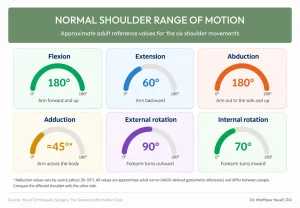
Understanding normal motion helps put restrictions in perspective. Approximate adult reference values for the shoulder are shown below (AAOS reference values).
| Movement | Description | Approximate normal range |
| Flexion | Arm raised forward and up | About 180° |
| Extension | Arm moved backward | About 60° |
| Abduction | Arm raised out to the side and up | About 180° |
| Adduction | Arm moved across the body | Roughly 30–50°, depending on the reference |
| External rotation | Forearm turns outward | About 90° |
| Internal rotation | Forearm turns inward | About 70° |
These figures vary between individuals. What matters clinically is comparing the affected shoulder with the other side and distinguishing active motion, which you produce yourself, from passive motion, which someone else produces for you. Loss of passive motion is the hallmark of frozen shoulder, while loss of active motion with preserved passive motion suggests a tendon or nerve problem.
When Anatomy Goes Wrong
Matching structures to common diagnoses helps make sense of a clinical explanation.
| Structure | Common problem |
| Rotator cuff tendons | Tendinopathy, partial or full tears |
| Subacromial bursa | Bursitis, impingement |
| Glenoid labrum | SLAP tears, Bankart lesions |
| Joint capsule | Frozen shoulder, instability |
| Articular cartilage | Osteoarthritis |
| Biceps tendon | Tendinitis, rupture |
| Acromioclavicular joint | AC separation, arthritis |
| Glenohumeral joint | Dislocation, instability |
At Yousif Orthopedic Surgery, a shoulder evaluation works systematically through these structures, since identifying which one is involved determines the whole treatment plan. Sports-related problems are covered in more detail on our sports injury page, and when non-surgical care is not enough, our shoulder surgery page explains the options.
When to See a Shoulder Specialist
Schedule an evaluation with an orthopedic specialist if shoulder pain lasts more than a few weeks, wakes you at night, or comes with weakness, catching, clicking, or a feeling that the shoulder is unstable.
Seek urgent medical care after a suspected dislocation (a visibly out-of-place shoulder or being unable to move the arm), sudden severe pain after an injury, numbness over the outer shoulder, or shoulder pain with fever or a hot, red, swollen joint. Chest pain, pressure, or shortness of breath with arm or shoulder pain is a medical emergency.
Reading a Shoulder Anatomy Diagram
If you are looking at a shoulder anatomy diagram for the first time, the labels can be overwhelming. A few orienting principles help. The anatomy is the same in the right and left shoulder, so a left shoulder diagram is simply a mirror image of a right one.
- Find the glenoid first. It is the shallow socket on the outer edge of the shoulder blade, and everything else is arranged around it.
- Identify the acromion. The bony roof above the joint defines the subacromial space beneath it.
- Trace the rotator cuff. These four muscles originate on the shoulder blade and wrap to attach on the humeral head.
- Note the labrum. It appears as a rim around the socket edge, easy to miss but functionally important.
- Look for the biceps tendon. It runs through its groove and blends into the top of the labrum.
Anterior views usually show the subscapularis and biceps tendon. Posterior views show the infraspinatus, teres minor, and the spine of the scapula. Recognizing which view you are looking at makes any diagram far easier to interpret.
Conclusion
Shoulder anatomy is a study in trade-offs. The shallow socket, loose capsule, and reliance on muscle for stability give the shoulder its extraordinary range of motion, and the same features explain its vulnerability to dislocation, tendon problems, and instability.
Knowing what sits inside the joint makes clinical explanations far easier to follow. The labrum deepens the socket, the capsule and ligaments provide passive restraint, the rotator cuff centers the ball actively, the bursae reduce friction, and the shoulder blade positions the entire system. When a clinician describes a labral tear, subacromial impingement, or a capsular problem, you now know which structure they mean and why it matters.
If shoulder pain or limited motion is affecting your daily life, the team at Yousif Orthopedic Surgery can identify which structure is involved and recommend the right treatment. Dr. Matthew Yousif, DO, is a fellowship-trained orthopedic surgeon with training in sports medicine and shoulder surgery. We see patients at our Troy and Shelby Township offices and serve Royal Oak, Southfield, Novi, Bloomfield Hills, and the greater Metro Detroit area. Explore our shoulder conditions or schedule an evaluation to get a clear answer.
References
- Anatomy, Shoulder and Upper Limb, Glenohumeral Joint. NIH StatPearls. https://www.ncbi.nlm.nih.gov/books/NBK537018/
- Anatomy, Shoulder and Upper Limb, Shoulder Muscles. NIH StatPearls. https://www.ncbi.nlm.nih.gov/books/NBK534836/
- Shoulder Joint Anatomy: Overview, Gross Anatomy, Microscopic Anatomy. Medscape. https://emedicine.medscape.com/article/1899211-overview
- The Shoulder Joint. Kenhub. https://www.kenhub.com/en/library/anatomy/the-shoulder-joint
- Rotator Cuff Injury. NIH StatPearls. https://www.ncbi.nlm.nih.gov/books/NBK547664/
6. American Academy of Orthopaedic Surgeons (AAOS) reference values for normal joint range of motion (goniometry).